Your Heart's Electrical Blueprint: Understanding the ECG/EKG
Published November 23, 2025
Have you ever seen those squiggly lines on a monitor in a hospital scene? That pattern is your Electrocardiogram (ECG or EKG)—one of the simplest, fastest, and most crucial tests doctors use to check the health of your heart. An ECG is essentially a picture of your heart's electrical activity. It helps doctors understand if your heart is beating properly, strong enough, and getting the blood flow it needs. The best part? It's painless, non-invasive, and useful for everyone, from newborns to the elderly.
How Does an ECG Work?
Your heart is a muscle, and like all muscles, it contracts thanks to tiny electrical impulses. These impulses start in the heart's natural pacemaker (the sinoatrial node) and spread through the muscle tissue, causing it to squeeze and pump blood. The ECG machine uses small, sticky sensors called electrodes placed on your chest, arms, and legs. These electrodes pick up the electrical signals that travel through your body. The machine then amplifies these signals and prints them out as a wave pattern on paper or displays them on a screen.
Interpreting the Waves
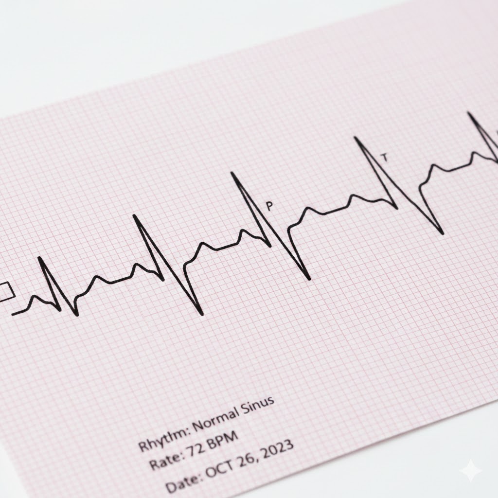
A healthy heart produces a predictable pattern, typically marked by P, QRS, and T waves.
- P wave: Shows the electrical activity starting in the upper chambers (atria), causing them to contract.
- QRS complex: Shows the electrical activity spreading to the lower, main pumping chambers (ventricles).
- T wave: Shows the electrical recharging (repolarization) of the ventricles, preparing for the next beat.
The Power of the ECG: What It Can Tell Your Doctor
The ECG provides a vast amount of information about the rhythm and structure of the heart. It's often the first step in diagnosing many heart conditions:
Arrhythmias: Irregular, too slow (bradycardia), or too fast (tachycardia) heart rhythms.
Heart Attack: Changes in the T and S-T waves that indicate heart muscle damage or lack of oxygen (ischemia).
Enlarged Heart: Larger-than-normal QRS complexes or certain wave changes that suggest the heart muscle has thickened.
Electrolyte Imbalance: Abnormal wave shapes caused by unusual levels of potassium, calcium, or other chemicals in the blood.
Pacemaker Function: Confirms that an implanted pacemaker is working correctly.
ECG in Adults: Detecting the Acute and Chronic
For adults, the ECG is an indispensable tool, especially in emergency and critical care.
Chest Pain: If you arrive at the emergency room with chest pain, an ECG is done immediately. It can quickly distinguish between dangerous heart attacks requiring immediate intervention and other causes of pain.
Screening: It's often used before surgery to ensure the heart can handle the stress of the procedure.
Monitoring Chronic Conditions: Patients with conditions like hypertension (high blood pressure) or atrial fibrillation (a common irregular heart rhythm) have regular ECGs to track changes over time and adjust medication.
ECG in Children:
Diagnosing Congenital Issues:
While adults often use the ECG to check for problems developed later in life, the ECG is essential in paediatrics for catching issues present since birth (congenital).
Heart Murmurs:
If a pediatrician hears an unusual heart sound (a murmur), an ECG helps determine if the sound is "innocent" (harmless) or caused by a structural defect, like a hole in the heart (Atrial or Ventricular Septal Defect).
Syncope (Fainting):
If a child faints, an ECG can rule out underlying electrical disorders, some of which are genetic and can be serious if untreated.
Congenital Heart Disease (CHD):
For children born with complex heart defects, the ECG monitors how the heart is coping with the increased strain or how it's recovering after surgical repair. The interpretation of a child's ECG is slightly different from an adult's, as a child's heart rhythm changes naturally as they grow, but its utility remains paramount.